 As, "necessity is the mother of invention", Canadian and other medical inventions are the reflections of particular circumstances: the need for vital diagnoses of the complex and very different illnesses and diseases. 245 babies are born every minute, and 102 people die each minute. If we do the math then 143 new members are added in the world population every minute. On Feb 24, 2010, 6:51, the world population was 6,804,833,483.
But how is it that the birth rate and the death rate are not equal? The answer is invention. The inventions have enabled the death rate to decrease and so every second the world population is different. But how do these medical inventions benefit people, i.e. humans? The most complex living thing on this planet is the human body. The inventions ensure our health, not just by helping doctors locate where and what disease we have, but also helping us live longer than we would by supplying to us, the supplements and medicine, that would help. These inventions also help us visualize this magnificent and unique living thing on earth.

This all did not just come from no where, but were great contribution of some remarkable people and entrepreneurs. The X-ray that came along in 1895, were discovered by Wilhelm Conrad Röntgen, who received the first Nobel Prize in Physics in 1901. As the November dusk darkened the laboratory, he had noticed that whenever he made sparks in the tube, a fluorescent screen at the other end of the laboratory table glowed slightly. This was the signal that he had been looking for, the sign that invisible rays were being produced in the spark tube, crossing the room and striking the screen, producing the faint glimmer. To track the rays he had been putting pieces of card in their way, but the screen continued to glow whether the cards were there or not as if the rays were able to pass clean through them. He then tried to block the rays with metal but thin pieces of copper and aluminium were as transparent as the card had been. He moved a piece of lead near to the screen, watching its shadow sharpen, and it was then that he dropped it in surprise: he had seen the dark skeletal pattern of the bones as his hand moved across the face of the screen. Still doubting what he saw he took out some photographic film for a permanent record. Röntgen had made one of t
 he most monumental discoveries in the history of science: X-rays, and seen for the first time images that are today common in every hospital casualty department. Six weeks later, on the Sunday before Christmas 1895, he invited his wife Bertha into the laboratory and took a shadow-graph of the bones of her hand with her wedding ring clearly visible. This is one of the most famous images in photographic history and propelled him within two more weeks into an international celebrity. The medical implications were immediately realised and the first images of fractured bones were being made by January 1896 even though none yet knew what the mystery rays were. (Nobleprize.org) This was one of the most important inventions as it set the basis for many other inventions.
One great inventions that came along after the X-ray was the CT or CAT scan. CT scan was discovered independently by a British engineer named Sir Godfrey Hounsfield and Dr. Alan Cormack. It has become a mainstay for diagnosing medical diseases. For their work, Hounsfield and Cormack were jointly awarded the Nobel Prize in 1979. CT scans are special x-ray tests that produce cross-sectional images of the body using x-rays and a computer. These images allow the radiologist, a medical doctor who specializes in images of the body, to look at the inside of the body just as you would look at the inside of a loaf of bread by slicing it. This type of special x-ray, in a sense, takes "pictures" of slices of the body so doctors can look right at the area of interest. CT scans are frequently used to evaluate the brain, spine, chest, abdomen, pelvis, and sinuses. Here's an article about how this development helps boost survival rates for severe injuries. A team of researchers led by Stefan Huber-Wagner of Munich University Hospital conducted the first large-scale study to find out whether the technique increased survival rates:
http://www.vancouversun.com/health/scans+boost+survival+rates+severe+injuries/1436028/story.html
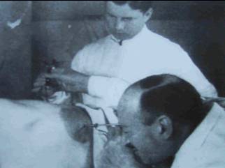
Another great invention that came that helps doctors and surgeons look at internal organs such as stomatch is endoscopy. Endoscope, the instrument used in endoscopy, is usually referred to as "an instrument used in medical operations which consists of a very small camera on a long thin tube which can be put into a person's body so
 that the parts inside can seen" (Oxford dictionary) or "An instrument for examining visually the interior of a bodily canal or a hollow organ such as the colon, bladder, or stomach." (Dictionary.com). Endoscopy is a medical operation in which an endoscope is put into a person's body so that the parts inside can be seen (Dictionary.com). In 1806, Philip Bozzini built an instrument that could be introduced in the human body to visualize the internal organs. He called this instrument "Lichtleiter". Bozzini used an aluminum tube to visualize the genitourinary tract. The tube, illuminated by a wax candle, had fitted mirrors to reflect images. In 1853, Antoine Jaen Desormeaux, a French surgeon first introduced the 'lichtleiter' of Bozzini to a patient. For many surgeons he is considered as the "Father of Endoscopy". Again in 1867, Desormeaux, used an open tube to examine the genitourinary tract, combining alsohol and turpentine with a flane in order to generate more condensable beam of light.
Another invention, that came as an innovation of the endoscopy, was the laparoscopy. Laparoscope, used in the surgery of laparosco
 py, is a slender tubular endoscope that is inserted through an incision in the abdominal wall and used for viewing the abdominal or pelvic cavities (Dictionary.com). Laparoscopy is an eximination of the inside of the body using a tube-shaped instrument that can be put through the wall of the abdomen. Laparoscopy, a branch of endoscopy, branched out after gastroscopy and hysteroscopy. The innovation, that came along the time, introduced laparoscopy in 1901, when the first experimental laparoscopy was performed in Berlin by a German surgeon Georg Kelling. Thus, it is difficult to credit one individual with the pioneering of the laparoscopic approach.
 First introduced to the medical world in 1950s, ultrasound has become the second most widely-used diagnostic imaging modality today. Sound waves of very high frequencies can easily and harmlessly penetrate human flesh. As waves enter the body, they encounter different materials such as the bone and internal organs. These materials cause the waves to reflect back to the source. Because the waves reflect back differently, physician can identify the type of tissue by the nature of the reflection.
The great discoveries in the medical world have lead to great decreases in the death rate and increases in the life expectancy rate. Using the latest technologies, doctors are more equipped and are more correct in their predictions about the diseases. These technologies have also enabled us to learn more about ore complex human bodies, and the internal body systems such as digestive, circulatory and respiratory, and have enabled doctors to find cure for their patients and perform successful surgeries.
Sources:
http://www.census.gov/main/www/popclock.html
http://www.wholesomewords.org/missions/greatc.html
http://www.discoveryofinsulin.com/Home.htm
http://www.vancouversun.com/health/scans+boost+survival+rates+severe+injuries/1436028/story.html
http://www.mta.ca/about_canada/study_guide/doctors/better_foods.html
http://inventors.about.com/od/mstartinventions/a/medicine.htm
http://nobelprize.org/educational_games/physics/x-rays/index.html
http://laparoscopy.blogs.com/endoscopyhistory/chapter_6/index.html
http://www.laparoscopyhospital.com/history_of_laparoscopy.htm
http://library.thinkquest.org/16541/eng/learn/library/content/ultrasound.htm
